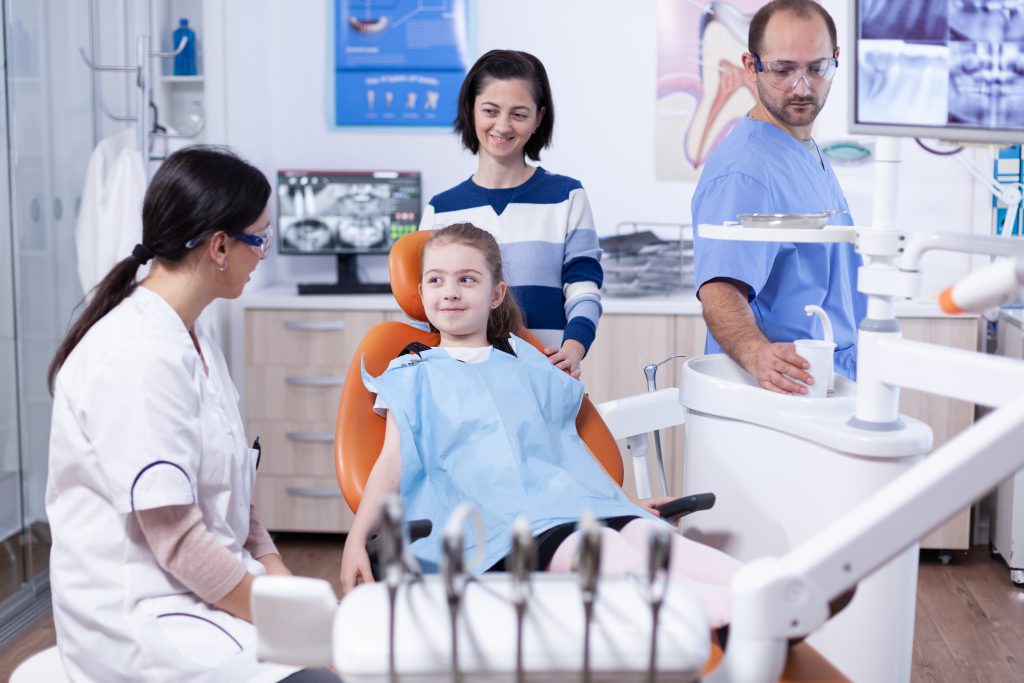
Common Procedures
A vast array of dental procedures may necessitate general anesthesia for patients. Below is a list of some of the most common ones.

Bonding is a conservative way to repair slightly chipped, discolored, or crooked teeth. During dental bonding, a white filling is placed onto your tooth to improve its appearance. The filling “bonds” with your tooth. Because it comes in a variety of tooth-colored shades, it closely matches the appearance of your natural teeth.
Tooth bonding (composite) can also be used for fillings instead of silver amalgam. Many patients prefer composite fillings because the white color is much less noticeable than silver. Composite fillings can be used on front or back teeth, depending on the location and extent of tooth decay.
Bonding is less expensive than other cosmetic treatments and can usually be completed in one visit to our office. However, bonding can stain and is easier to break than other cosmetic treatments, such as porcelain veneers. If it breaks or chips, let us know. The bonding can generally be patched or repaired easily during a single visit.
Crowns are a restorative procedure used to improve your tooth’s shape or to strengthen it. Crowns are most often used for teeth that are broken, worn, or have portions destroyed by tooth decay.
A crown is a “cap” cemented onto an existing tooth that usually covers the portion of your tooth above the gum line. In effect, the crown becomes your tooth’s new outer surface. Crowns can be made of porcelain, metal, or both. Porcelain crowns are most often preferred because they mimic the translucency of natural teeth and are very strong.
Crowns or onlays (partial crowns) are needed when there is insufficient tooth strength to hold a filling. Unlike fillings, which apply the restorative material directly into your mouth, a crown is fabricated away from your mouth.
Your crown is created in a lab from your unique tooth impression, which allows a dental laboratory technician to examine all aspects of your bite and jaw movements. Your crown is sculpted just for you so your bite and jaw movements function normally once the crown is placed.
There are times when it is necessary to remove a tooth. Sometimes a baby tooth has misshapen or long roots that prevent it from falling out as it should, and the tooth must be removed to make way for the permanent tooth to erupt.
At other times, a tooth may have so much decay that it puts the surrounding teeth at risk, so our team may recommend removal and replacement with a bridge or implant. Infection, orthodontic correction, or problems with a wisdom tooth can also require removal of a tooth.
When it is determined that a tooth needs to be removed, your doctor may extract it during a regular checkup or request another visit for this procedure. The root of each tooth is encased within your jawbone in a “tooth socket,” and your tooth is held in that socket by a ligament. In order to extract a tooth, the doctor must expand the socket and separate the tooth from the ligament holding it in place. While this procedure is typically very quick, it is important to share with us any of your concerns or preferences for sedation.
Once a tooth has been removed, neighboring teeth may shift, causing problems with chewing or your jaw joint function. To avoid these complications, we may recommend you replace the extracted tooth.
Traditional dental restoratives or fillings are most often made of silver amalgam. The strength and durability of this traditional dental material makes it useful for situations where restored teeth must withstand extreme forces that result from chewing, often in the back of the mouth.
Newer dental fillings include ceramic and plastic compounds that mimic the appearance of natural teeth. These compounds, often called composite resins, are usually used on the front teeth where a natural appearance is important, but they can also be used on the back teeth, depending on the location and extent of the tooth decay.
There are two different kinds of fillings: direct and indirect. Direct fillings are placed into a prepared cavity during a single visit. They include silver amalgam, glass ionomers, resin ionomers, and composite (resin) fillings.
Indirect fillings generally require two or more visits. They include inlays, onlays, and veneers. They are used when a tooth has too much damage to support a filling, but not enough to necessitate a crown.
If your child’s primary tooth has extensive decay, or has been damaged by trauma, action may be needed to restore the integrity of the tooth and prevent infection from spreading to surrounding teeth. After a set of X-rays are taken, we will be able to assess the extent of the infection and recommend one of two options: a pulpotomy or a pulpectomy.
PULPOTOMY
If the decay or trauma is confined to the crown of the tooth, a pulpotomy may be recommended. When a cavity gets really deep, close to the pulp of a tooth, or even into the pulp, the pulpal tissue becomes irritated and inflamed.
A pulpotomy is when the inflamed pulp chamber, usually on a baby molar, is removed. The doctor will remove all the infected material in the pulp of the crown only, but leave the living tooth root intact. After a pulpotomy on a baby molar, the empty space will be filled with dental cement and a stainless steel crown will be placed to restore the tooth.
PULPECTOMY
If the infection involves tissue in both the tooth crown and the tooth root, a pulpectomy may be the best option. In a pulpectomy, the entire pulp material is removed from both the crown and the roots. After numbing your child’s tooth, the dentist will remove the pulp and nerve tissue from the crown and from the canals of the roots.
Then the pulp chamber and root canals will be thoroughly cleaned and disinfected. Next, the doctor will fill the tooth and tooth roots with a dental cement, and finish with a stainless steel crown.
CROWNS
Crowns are “cemented” onto an existing tooth and fully cover the portion of the tooth above the gum line. In effect, the crown becomes the tooth’s new outer surface.
Stainless steel dental crowns are used as a good temporary restoration to save a primary tooth until the permanent tooth can erupt and take its place. Keeping the primary tooth, if at all possible, is desirable.
A primary tooth can be restored with a stainless steel crown during one appointment. A crowned tooth must be brushed and flossed just like other teeth.
In the past, if you had a tooth with a diseased nerve, you’d probably lose that tooth. Today, with a special dental procedure called “root canal treatment,” your tooth can be saved.
When a tooth is cracked or has a deep cavity, bacteria can enter the pulp tissue and germs can cause an infection inside. If left untreated, an abscess may form.
If the infected tissue is not removed, pain and swelling can result. This can not only injure your jawbones, but it is also detrimental to your overall health.
Root canal treatment involves one to three visits. During treatment, a general dentist or endodontist (a dentist who specializes in problems with the nerves of the teeth) removes the affected tissue. Next, the interior of the tooth will be cleaned and sealed.
Finally, the tooth is filled with a dental composite. If the tooth has extensive decay, the doctor may suggest placing a crown to strengthen and protect the tooth from breaking. As long as you continue to care for your teeth and gums with regular brushing, flossing, and checkups, your restored tooth can last a lifetime.
If your child’s tooth has come out too soon because of decay or an accident, it is crucial to maintain the space to prevent future space loss and dental problems when permanent teeth begin to come in. Without the use of a space maintainer, the teeth that surround the open space can shift, and impede the permanent tooth’s eruption. When that happens, the need for orthodontic treatment may become greater.
TYPES OF SPACE MAINTAINERS
Space maintainers can be made of stainless steel and/or plastic, and can be removable or fixed (cemented to the teeth).
REMOVABLE
A removable space maintainer looks much like a retainer with plastic blocks to fill in where the tooth is missing. If your child is older and can reliably follow directions, a removable space maintainer can be a good option.
FIXED
Fixed space maintainers come in many designs.
A band-and-loop maintainer is made of stainless steel wire and held in place by a crown or band on the tooth adjacent to the empty space. The wire is attached to the crown or loop and rests against the side of the tooth on the other end of the space.
A lingual arch is used on the lower teeth when the back teeth on both sides of the jaw are lost. A wire is placed on the lingual (tongue) side of the arch and is attached to the tooth in front of the open space on both sides. This prevents the front teeth from shifting backwards into the gap.
In the case of a lost second primary molar prior to the eruption of the first permanent molar, a distal shoe may be recommended. Because the first permanent molar has not come in yet, there is no tooth to hold a band-and-loop space maintainer in place. A distal shoe appliance has a metal wire that is inserted slightly under the gum and will prevent the space from closing.
CARING FOR YOUR CHILD’S SPACE MAINTAINER
There are four general rules for taking care of your child’s appliance.
- Your youngster should avoid sticky foods, including candy and chewing gum.
- Encourage your son or daughter not to push or tug on the space maintainer with the fingers or tongue.
- Keep your child’s space maintainer clean through effective brushing and flossing.
- Your little one should continue to see the pediatric dentist for regular dental visits.
Tongue-tie is a birth defect that occurs when the strip of skin (lingual frenulum) that connects a baby’s tongue to the floor of the mouth is shorter than usual. Typically, this strip of skin separates before birth, which allows the tongue free range of motion. With tongue-tie, the lingual frenulum remains attached to the bottom of the tongue.
Tongue-tie is a common condition that, if addressed quickly, will not hinder a child’s development. However, if left untreated, tongue-tie can result in malnourishment, speech difficulty, or poor oral hygiene.
SIGNS OF TONGUE-TIE INCLUDE:
- Restriction of the tongue’s movement, making it harder to breastfeed.
- Difficulty lifting the tongue up or moving it from side to side.
- Difficulty sticking the tongue out.
- The tongue looks notched or heart-shaped when stuck out.
TREATMENT OF TONGUE-TIE
The treatment of tongue-tie for infants is a simple surgical procedure called a frenectomy. A pediatric specialist examines the lingual frenulum and then uses sterile scissors or laser to snip the frenulum free. Stitches are usually not necessary. Since there are few nerve endings or blood vessels in the lingual frenulum, only a local anesthetic is used.
Frenotomy for tongue-tie in older children and adults is similar to that for infants, although it is usually done under general anesthesia and may involve stitches. Speech therapy may also be necessary.